Total anomalous pulmonary venous correction in India
TAPVC is a congenital heart disease. It is an anomaly affecting the blood flow from pulmonary veins to the heart
What is Total anomalous pulmonary venous correction?
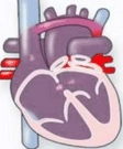
Total Anomalous Pulmonary Venous Connection (TAPVC) is a type of congenital heart disease (CHD). Here, there is an ‘anomaly’ or abnormality in the way pulmonary veins empty into the heart.
TAPVC constitutes 2-3% of all CHDs. In TAPVC, the veins carrying oxygen-rich blood to the heart from the lungs, empty into the right atrium instead of the left. It reduces the level of oxygen in the blood. Hence, babies with TAPVC have a bluish tint to their skin.
Breathlessness, rapid heart rate, failure to gain weight, repeated chest infections are other common symptoms. In addition to this, there is a hole between the two upper chambers of the heart (atrial septal defect).
An open heart surgery named TAPVC repair surgery can correct this defect. The surgeon shifts the attachment of the pulmonary veins to the left atrium from the right and closes the hole. This surgery restores the normal circulation path in the heart.
A pediatric cardiac surgeon does the surgery. The duration of the operation is around 3 to 5 hours. Your baby will be in the hospital for 10-12 days.
If not treated, only 50 % of children survive beyond three months, and only 20% survive beyond their first birthday. Those neonates who have an obstruction in the pulmonary venous pathway may need emergency surgery within a few hours to a few days of life. Others who are stable can undergo surgery between 3-4 months.
What is TAPVC?
In a normal heart, the oxygen-poor blood comes into the right atrium through two big veins, the superior and inferior vena cava. From the right atrium, this blood flows to the right ventricle. The pulmonary arteries take this blood to the lungs for oxygenation.
The oxygen-rich blood is then taken out of the lungs by the pulmonary veins to the left atrium. From the left atrium, the blood enters the left ventricle, which then pumps it into the aorta. The aorta then branches out to supply various parts of the body with oxygen-rich blood.
The pulmonary veins of a baby with TAPVC drains into the right atrium instead of the left atrium. So, the left ventricle will not receive enough blood to pump into the aorta creating problems with blood circulation.
To compensate, most babies with a TAPVC will have a hole in the wall between the right and left atrium, called an atrial septal defect (ASD). This hole allows the flow of blood from the right to the left atrium.
But the blood moving from the right atrium to the left will be a mixture of oxygen-rich and oxygen-poor blood. So, a baby with a TAPVC will have low levels of oxygen in the blood.
Few babies with TAPVC will have a block in the pulmonary veins leading to a build-up of oxygen-rich blood in the lungs. This condition is called pulmonary hypertension, which damages the lungs as well. These children will need emergency surgery.
Types of TAPVC
There are four types of TAPVC classified according to the way the pulmonary veins drain into the right atrium. These are as follows;
- Supracardiac TAPVC – The pulmonary veins drain into the right atrium through the superior vena cava vein(SVC). It is a large vein that brings deoxygenated blood from the upper part of the body to the heart. It is the most common type of TAPVC seen in around 45% of cases.
- Infra-cardiac TAPVC – The pulmonary veins are attached to the hepatic vein and the inferior vena cava vein(IVC). The hepatic vein brings deoxygenated blood from the liver and the inferior vena cava from the lower part of the body. Seen in 25% of TAPVC cases, it is the most common type to have a pulmonary venous obstruction. It leads to severe pulmonary hypertension requiring emergency surgery.
- Cardiac TAPVC – The pulmonary veins drain directly into the right atrium or the coronary sinus. Coronary sinus brings blood from the heart muscles into the right atrium.
- Mixed TAPVC – It is a combination of the above types. About 5-10% of the total cases are mixed TAPVC cases. Right pulmonary veins drain into SVC and left pulmonary veins into the hepatic vein and IVC.
All these types of TAPVC have an atrial septal defect (ASD) associated with it, and it is essential for survival. ASD is an opening in the wall that separates the right and left atrium. It enables blood to move over to the left atrium and then to the body via the left ventricle.
The size of the ASD determines the clinical picture. If the ASD is small, the baby will need urgent surgery.
Partial anomalous pulmonary venous return
In this condition, one or two of the pulmonary veins will drain to the left atrium, and the others will drain into the right atrium. This condition is not as severe as a TAPVC, because a good amount of oxygen-rich blood reaches the left atrium.
Symptoms of TAPVC
The most common symptoms are
- Difficulty in breathing
- Poor feeding
- Poor weight gain
- Repeated chest infections
- Blue color in the lips, skin, etc
A doctor can hear a murmur or a ‘whoosh’ sound while using a stethoscope. It is due to the abnormal flow of blood from the right to the left atrium across the ASD.
TAPVC Diagnosis
There are various ways to diagnose TAPVC.
- Echocardiography - This test confirms the presence of TAPVC. It will help the doctor in assessing any abnormality in the size of the heart, the size of the ASD, and the volume of blood passing through it. It can help in finding out the type of TAPVC as well.
- Cardiac catheterization – While not always required, it will show how the blood flows through the pulmonary veins to the heart. The doctor can see if there are any obstructions in blood flow.
- Chest X-ray - It will show if there is an enlargement of the heart and the pulmonary artery. If an obstruction is present, the X-ray will show signs of fluid accumulation (edema) in the lungs.
- Abnormal heart sounds- There will be a heart murmur due to the flow of the blood from the right to the left atrium.
- Oxygen level in the blood - The levels of oxygen will be low in the blood of babies with TAPVC.
Treatment for TAPVC
TAPVC repair surgery is the only treatment option for a TAPVC.
In babies with poor health and who are not fit for open-heart surgery, but not having any venous obstruction, a cardiac catheterization procedure increases the size of the ASD. It increases the flow of blood to the left atrium and will help the baby until the surgery.
The baby will be under general anesthesia during the TAPVC surgery. Some babies will require life support with ECMO during and soon after surgery.
During the operation, the surgeon will attach the pulmonary veins into its natural position, which is the left atrium. The surgeon will also remove any obstructions in the pulmonary veins. All other routes of blood flow from the pulmonary veins to other veins will be tied up.
After surgery, the blood will flow from the lungs to the heart through the pulmonary veins, only into the left atrium. The next step in operation will be the closure of the ASD to prevent the abnormal blood flow from the right to the left atrium.
The surgery will take about 3 to 4 hours. Your baby will be in the hospital for about ten to twelve days.
Risks and complications of TAPVC surgery
TAPVC surgery has a very high rate of success and less chance of complications.Some of the babies may develop certain complications later on in their lives. The occurrence of pulmonary vein obstruction is one of the most common ones. They will have to go through a procedure like a balloon dilatation or another surgery then.
Chances of complications are more for babies who have to undergo emergency TAPVR surgery. These babies usually have severe obstructions in their pulmonary veins that make them very weak, and there will be high pressure in the lungs. This condition is pulmonary hypertension. They will be on external life support like ECMO before, during, and for some time after surgery.
Recovery from a TAPVC surgery
After the surgery, the baby will be shifted to a pediatric cardiac intensive care unit(PCICU) and will remain there for 5-6 days. While in the PCICU, the baby will have wires attached to the body to monitor the heart and body functions. There will also be tubes coming out from the chest to drain the blood collected inside and in the urinary system to monitor the urine output.
The doctors will use an echocardiogram to assess the functioning of the heart. Very rarely, external life support may be necessary to lessen the burden on the heart and lungs. It also helps in faster recovery.
The baby is taken off the ventilator support once he is awake and hemodynamically stable with no bleeding from the chest tubes. The medication will be gradually decreased and stopped over the next 3-4 days.
The baby will be shifted to the room after 2 or 3 days, depending on the progress. Even when in the room, wires and leads will be attached to the baby’s body to monitor the heart function.
If everything goes on as planned, you can go back along with the baby after 10 to 12 days. The doctor will give you steps on how to care for the baby at home and about the follow-up visits. The follow-up visits will help assess the progress of the baby and the speed of recovery.
Top Hospitals for Total anomalous pulmonary venous correction
We partner with India's most respected accredited hospitals.


Frequently Asked Questions
Total anomalous pulmonary venous connection(Tapvc) or Total anomalous pulmonary venous return(Tapvr) is a congenital heart defect
It is an anomaly affecting the blood flow from pulmonary veins to the heart. Here, the veins that carry blood from the lungs to the heart flow into the right atrium instead of the left atrium. It causes a reduction in the level of oxygen in the blood. Tapvr surgery is the procedure to correct this problem.
The prevalence of Tapvr is about 1 in 10,000 births
It is a rare type of congenital heart disease(CHD), making up about 2% of the total CHDs. However, it is a severe case that needs medical attention and surgery
There are four main types of Tapvc, out of which Supracardiac Tapvc is the most common.
The four types are
- Supracardiac Tapvc which accounts for more than 45% of all cases
- Infracardiac Tapvc contributing to 25% of all cases
- Cardiac Tapvc and
- Mixed Tapvc
Get a Free Quote
We respond within 2 working hours.
Request Received!
A coordinator will contact you within 2 working hours.