Retinal Detachment in India
Retinal Detachment is an emergency eye medical condition where the retina gets detached from the tissues supporting it
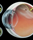
What is Retinal Detachment?
Retinal detachment happens when the retina separates from the back wall of your eye. The retina is the light-sensitive tissue at the back of your eye where images form. The optic nerve transfers these images to the brain to create the sense of vision.
Your retina needs a constant supply of blood to help it function properly. If you have a detached retina, there will be a decrease in blood flow that can cause damage. You may lose a part or whole of your sight due to retinal detachment. It will depend on the extent of the detachment you have.
The blurring of vision, floaters, loss of side vision, sudden flashes of light, etc. are some of the early symptoms. You will need early treatment to prevent the condition from getting worse. Delay in treatment will lead to a total loss of vision.
There are different types of retinal detachments according to its cause. Leakage of fluid inside the retina, scar formation, etc. are some of the common causes.
There are different treatments for retinal detachment according to cause and your condition. The goal of all treatments is to reattach the retina to its place. You will also have treatment to remove the cause as well.
Causes and types of retinal detachment
There are three main types of retinal detachment. The causes for these are also different. The types and their causes are;
- Rhegmatogenous.- It is the most common type. The cause is usually a small tear in the retina. The vitreous humor present inside your eyeball slowly starts seeping inside the retina through the hole. After some time, the fluid slowly starts to put pressure on the retina to separate it from the back of the eye. Eye injury, eye surgery, etc. are some of the reasons for a retinal tear.
- Tractional - You will have this if you have severe diabetes that leads to retinopathy. In this, there will be bleeding from the blood vessels in the retina. It results in scars that may pull the retina away from the back of the eye.
- Exudative- It happens when fluids start forming behind the retina due to inflammation or eye injury. This fluid slowly starts putting pressure on the retina to move it away from the back of the eye.
Risk factors of retinal detachment
A few things can increase your chance of having a retinal detachment. Some of them are
- Having severe short-sight or nearsightedness- You are at high risk as it causes a lot of strain to the eyes
- Having family members with the disease- Your risk is more as it runs in families
- Your age- Your risk will be more if you are above sixty
- Trauma to the eye- The risk is more if you have had an eye injury
- History of retinal detachment before- You are more likely to have it again
- Having severe or uncontrolled diabetes- If your blood sugar levels are high for a long time, your risk is more
- Cataract surgery- Some people may develop retinal detachment after it
You may have the condition without any of these risk factors. Also, not everybody with risk factors gets it as well.
Symptoms of retinal detachment
Some of the symptoms of retinal detachment will include;
- Seeing several 'floaters' before your eyes - Floaters are dark spots that appear as specks or streaks in front of your eyes. In some, it may occur without any reason. But if you see many large floaters all on a sudden, it may be an indication of retinal detachment
- Sudden flashes of light- It is one of the early signs. You will see sudden flashes of light, especially from the sides.
- Dark shadows in your field of vision- You will see dark shadows on the side of your field of vision
- A moving dark curtain on your field of vision- You will see a dark curtain moving across your field of vision.
- A sudden decrease in your sight
- Partial loss of sight starting from the sides
- Total loss of vision
In a few, retinal detachment may occur all of a sudden without any warning signs.
Diagnosis of retinal detachment
If you have any signs of retinal detachment, you will have the following tests.
- Retinal examination - During this, the doctor will look into your retina with the help of a microscope and light. It helps to detect any tears, holes, or cracks in the retina. The doctor will also have an idea about the extent of retinal detachment.
- Ultrasound eye scans- You will have this if there is blood in the eyes that prevents the doctors from looking into the retina. The scans will show the extent of damage and the area of tears.
Treatments for retinal detachment
Your treatment will depend on the extent of retinal detachment.
If it is small, you will have
- Laser surgery or photocoagulation - This procedure uses lasers to make small burns around the tear. It will result in scars that help attach the retina to the eyewall
- Cryopexy- It uses a freezing probe to make small and delicate scars in the retina. The scars help attach the retina back to the eyewall.
Surgery for retinal detachment
You will need surgery to repair large retinal detachments. The type of surgery you will have will depend on the cause and the extent of the damage.
Pneumatic retinopexy
You will have this treatment if
- A single tear or break is the cause of your detachment
- The tear is in the upper part of the retina
- You have multiple tears in the retina, but they are close to each other
Before the procedure, your doctor will apply a local anesthetic to make your eye numb. After this, the doctor will inject a gas bubble into your eyeball. The bubble will press the retina against the back of your eye.
The doctor will then use lasers or a freezing probe to seal the tear or hole in the retina. It prevents the condition from coming back.
During it, you will have to hold your head in a position where the bubble and the tear are at its highest point. That is why the procedure is not effective in tears that are at the bottom of the retina. In such cases, you will have to keep your head upside down to keep the bubble in position.
Scleral buckling
It is a procedure to treat rhegmatogenous retinal detachments. In this, your doctor will place a silicone or sponge buckle in the sclera of your eyes. The buckle will push the sclera towards the retinal tear and make the retina flatter. It makes the retina push close to your eye's inner wall and helps in attaching to it again. Scleral buckling thus helps to prevent the tear from occurring again.
You will have the procedure under local or general anesthesia. The doctor will place the buckle close to the retinal tear by making a small cut in the sclera. The next step is to suture the buckle closely to prevent it from getting loose.
The surgeon will use laser or freezing probes to close the tear by making scars in the area. The scars seal the tears and prevent the vitreous fluid from seeping below the retina.
It is a safe procedure with very few risks and complications. You will have to be in the hospital for only a day.
Vitrectomy
You will have this procedure if the detachment is due to the pull of the vitreous fluid on the retina.
During the procedure, the surgeon will drain the vitreous fluid along with the blood clots. For this, the doctor will make a small cut in the sclera. The surgeon will then use lasers or freezing probes to close the tears. After this, the surgeon will fill up the eyeball with a gas bubble or fluids like saline or silicone oil. The gas or saline will dissolve slowly move into the bloodstream, and natural fluid will start filling up the space. If you have silicone oil, you will need a small operation after a few months to remove it.
Prognosis
Most of those who have surgery have excellent results. The results will depend on the extent of detachment and the cause for it. In a few, the outcome will not be very favorable.
After surgery, the eyesight will improve to a great extent than before. It will also help to prevent the problem from coming back.
Top Hospitals for Retinal Detachment
We partner with India's most respected accredited hospitals.


Frequently Asked Questions
The risk factors for retinal detachment include;
- Having severe short-sight or nearsightedness
- Having family members with the disease
- Age above sixty
- History of eye injury, previous retinal detachment, cataract surgery
- Having severe or uncontrolled diabetes for a long time
Retinal detachment has very few early symptoms. You will have symptoms when the condition worsens. These include
- Seeing several 'floaters' or dark spots before your eyes
- Sudden flashes of light, especially from the sides
- Dark shadows or having a moving dark curtain in your field of vision
- Partial loss of sight starting from the sides
- Total loss of vision
In a few, retinal detachment may occur suddenly and without warning signs
If the detachment is small, you will have treatments like;
- Laser surgery or photocoagulation that uses lasers to make small burns to create scars that help attach the retina to the eyewall
- Cryopexy which uses a freezing probe to make small and delicate scars in the retina that help attach the retina back to the eyewall.
Your doctor will decide on the treatment by assessing the extent of detachment
If the detachment is severe you will have;
- Pneumatic retinopexywhere the doctor injects a gas bubble into the eyes to push and keep the retina against the eye wall
- Scleral buckling where the doctors attach small buckles in the white of the eye to put pressure on the retina to move back
- Vitrectomy to remove the vitreous fluid tif it tugs and pulls the retina from the eye wall
Get a Free Quote
We respond within 2 working hours.
Request Received!
A coordinator will contact you within 2 working hours.